Application of Riboflavin and Curcumin as Natural Photosensitizers for Antimicrobial Photodynamic Inactivation against Staphylococcus aureus: A Study for Skin Disease
DOI:
https://doi.org/10.59796/jcst.V15N4.2025.143Keywords:
curcumin, photodynamic inactivation, riboflavin, skin disease, Staphylococcus aureusAbstract
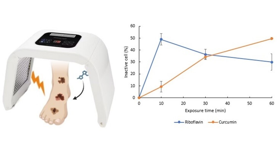
Atopic Dermatitis (AD) and Actinic Keratosis (AK) are becoming increasingly prevalent in developing countries, including Indonesia. Staphylococcus aureus, a common microorganism associated with both conditions, presents treatment challenges due to the increasing antibiotic resistance and associated side effects. Photodynamic Inactivation (PDI), which employs natural photosensitizers such as riboflavin and curcumin in combination with Omega Light LED-a technology commonly used in aesthetic treatments-offers a potential alternative. In this study, riboflavin and curcumin were applied separately at a concentration of 0.015% (w/v) and irradiated with red (λ = 640 nm), blue (λ = 423 nm), or green (λ = 532 nm) light using an Omega Light LED device (O'melon). Cell viability was assessed using an ELISA reader at 595 nm after irradiation durations of 10, 30, and 60 minutes. Skin toxicity was predicted using Toxtree 3.1.0, Pred-Skin 3.0, and pkCSM web-based tools. Results showed that the photosensitizers without irradiation were not cytotoxic to Staphylococcus aureus. However, the combination of blue light and photosensitizers significantly inhibited bacterial viability. Riboflavin achieved 49.0±4.8% inhibition within 10 minutes, indicating a rapid but transient effect, whereas curcumin elicited a slower yet sustained antibacterial response, achieving 34.2 ± 1.6% inhibition after 30 minutes. Computational toxicity predictions suggested no clear evidence of skin irritation; however, a potential for skin sensitization remains. These findings support the potential of riboflavin- and curcumin-based PDI using Omega Light LED as a promising non-antibiotic approach for managing Staphylococcus aureus infections in AD and AK.
References
Ablon, G. (2018). Phototherapy with light emitting diodes: Treating a broad range of medical and aesthetic conditions in dermatology. Journal of Clinical and Aesthetic Dermatology, 11(2), 21–27.
Almenara-Blasco, M., Pérez-Laguna, V., Navarro-Bielsa, A., Gracia-Cazaña, T., & Gilaberte, Y. (2024). Antimicrobial photodynamic therapy for dermatological infections: Current insights and future prospects. Frontiers in Photobiology, 2, Article 1294511. https://doi.org/10.3389/fphbi.2024.1294511
Alves, V. M., Capuzzi, S. J., Braga, R. C., Borba, J. V., Silva, A. C., Luechtefeld, T., ... & Tropsha, A. (2018). A perspective and a new integrated computational strategy for skin sensitization assessment. ACS Sustainable Chemistry & Engineering, 6(3), 2845-2859. https://doi.org/10.1021/acssuschemeng.7b04220
Alves, V. M., Capuzzi, S. J., Muratov, E. N., Braga, R. C., Thornton, T. E., Fourches, D., ... & Tropsha, A. (2016). QSAR models of human data can enrich or replace LLNA testing for human skin sensitization. Green Chemistry, 18(24), 6501-6515. https://doi.org/10.1039/C6GC01836J
Borba, J. V., Braga, R. C., Alves, V. M., Muratov, E. N., Kleinstreuer, N., Tropsha, A., & Andrade, C. H. (2020). Pred-skin: A web portal for accurate prediction of human skin sensitizers. Chemical Research in Toxicology, 34(2), 258-267. https://doi.org/10.1021/acs.chemrestox.0c00186
Borgia, F., Li Pomi, F., Vaccaro, M., Alessandrello, C., Papa, V., & Gangemi, S. (2022). Oxidative stress and phototherapy in atopic dermatitis: Mechanisms, role, and future perspectives. Biomolecules, 12(12), Article 1904. https://doi.org/10.3390/biom12121904
Braga, R. C., Alves, V. M., Muratov, E. N., Strickland, J., Kleinstreuer, N., Trospsha, A., & Andrade, C. H. (2017). Pred-skin: A fast and reliable web application to assess skin sensitization effect of chemicals. Journal of Chemical Information and Modeling, 57(5), 1013-1017. https://doi.org/10.1021/acs.jcim.7b00194
Bylund, S., von Kobyletzki, L. B., Svalstedt, M., & Svensson, Å. (2020). Prevalence and incidence of atopic dermatitis: A systematic review. Acta Dermato-venereologica, 100(12), Article 5765. https://doi.org/10.2340/00015555-3510
Chacon, J. N., McLearie, J., & Sinclair, R. S. (1988). Singlet oxygen yields and radical contributions in the dye‐sensitised photo‐oxidation in methanol of esters of polyunsaturated fatty acids (oleic, linoleic, linolenic and arachidonic). Photochemistry and Photobiology, 47(5), 647-656. https://doi.org/10.1111/j.1751-1097.1988.tb02760.x
Chaudhari, S. P., Tam, A. Y., & Barr, J. A. (2015). Curcumin: A contact allergen. The Journal of Clinical and Aesthetic Dermatology, 8(11), 43-48.
Chignell, C. F., Bilski, P., Reszka, K. J., Motten, A. G., Sik, R. H., & Dahl, T. A. (1994). Spectral and photochemical properties of curcumin. Photochemistry and Photobiology, 59(3), 295-302. https://doi.org/10.1111/j.1751-1097.1994.tb05037.x
Correia, J. H., Rodrigues, J. A., Pimenta, S., Dong, T., & Yang, Z. (2021). Photodynamic therapy review: Principles, photosensitizers, applications, and future directions. Pharmaceutics, 13(9), Article 1332. https://doi.org/10.3390/pharmaceutics13091332
Crusca, J. D. S., de Moraes, L. H. O., Figueira, T. G., Parizotto, N. A., & Rodrigues, G. J. (2025). Photodynamic therapy effects with curcuma longa l. active ingredients in gel and blue led on acne: A randomized, controlled, and double-blind clinical study. Photonics, 12(1), Article 80. https://doi.org/10.3390/photonics12010080
Dai, T., Gupta, A., Huang, Y. Y., Sherwood, M. E., Murray, C. K., Vrahas, M. S., ... & Hamblin, M. R. (2013). Blue light eliminates community-acquired methicillin-resistant Staphylococcus aureus in infected mouse skin abrasions. Photomedicine and Laser Surgery, 31(11), 531-538. https://doi.org/10.1089/pho.2012.3365
de Oliveira, E. F., Tikekar, R., & Nitin, N. (2018). Combination of aerosolized curcumin and UV-A light for the inactivation of bacteria on fresh produce surfaces. Food Research International, 114, 133-139. https://doi.org/10.1016/j.foodres.2018.07.054
Djalil, A. D., Susanto, A. P., Arisugita, R. N., Dhiani, B. A., Maulidan, M. F., & Zamzani, I. (2023). Natural dyes as photosensitizers of propionibacterium acnes [Conference presentation]. Proceedings of the Conference on Natural Resources and Life Sciences 2022 (NRLS-BIO 2022), Surabaya, Indonesia. https://doi.org/10.2991/978-94-6463-322-1_20
EFSA Panel on Additives and Products or Substances used in Animal Feed (EFSA FEEDAP Panel), Rychen, G., Aquilina, G., Azimonti, G., Bampidis, V., Bastos, M. D. L., ... & Wallace, R. J. (2018). Safety and efficacy of vitamin B2 (riboflavin) produced by Ashbya gossypii DSM 23096 for all animal species based on a dossier submitted by BASF SE. EFSA Journal, 16(7), Article e05337. https://doi.org/10.2903/j.efsa.2018.5337
Fekrirad, Z., Kashef, N., & Arefian, E. (2019). Photodynamic inactivation diminishes quorum sensing-mediated virulence factor production and biofilm formation of Serratia marcescens. World Journal of Microbiology and Biotechnology, 35(12), Article 191. https://doi.org/10.1007/s11274-019-2768-9
Ferreira dos Santos, R., Campos, B. S., Filho, F. D. A. M. R., de Oliveira Moraes, J., Albuquerque, A. L. I., Delgado da Silva, M. C., ... & de Araujo, M. T. (2019). Photodynamic inactivation of S. aureus with a water-soluble curcumin salt and an application to cheese decontamination. Photochemical & Photobiological Sciences, 18(11), 2707-2716. https://doi.org/10.1039/c9pp00196d
Galati, L., Brancaccio, R. N., Robitaille, A., Cuenin, C., Luzi, F., Fiorucci, G., ... & Tommasino, M. (2020). Detection of human papillomaviruses in paired healthy skin and actinic keratosis by next generation sequencing. Papillomavirus Research, 9, Article 100196. https://doi.org/10.1016/j.pvr.2020.100196
George, C. D., Lee, T., Hollestein, L. M., Asgari, M. M., & Nijsten, T. (2024). Global epidemiology of actinic keratosis in the general population: A systematic review and meta-analysis. British Journal of Dermatology, 190(4), 465-476. https://doi.org/10.1093/bjd/ljad371
Glass, G. E. (2021). Photobiomodulation: The clinical applications of low-level light therapy. Aesthetic Surgery Journal, 41(6), 723-738. https://doi.org/10.1093/asj/sjab025
Glatz, M., Bosshard, P. P., Hoetzenecker, W., & Schmid-Grendelmeier, P. (2015). The role of Malassezia spp. in atopic dermatitis. Journal of Clinical Medicine, 4(6), 1217-1228. https://doi.org/10.3390/jcm4061217
Golden, E., Ukaegbu, D. C., Ranslow, P., Brown, R. H., Hartung, T., & Maertens, A. (2023). The good, the bad, and the perplexing: Structural alerts and read-across for predicting skin sensitization using human data. Chemical Research in Toxicology, 36(5), 734-746. https://doi.org/10.1021/acs.chemrestox.2c00383
Gomez, C., Muangnoi, C., Sorasitthiyanukarn, F. N., Wongpiyabovorn, J., Rojsitthisak, P., & Rojsitthisak, P. (2019). Synergistic effects of photo-irradiation and curcumin-chitosan/alginate nanoparticles on tumor necrosis factor-alpha-induced psoriasis-like proliferation of keratinocytes. Molecules, 24(7), Article 1388. https://doi.org/10.3390/molecules24071388
Górnicka, J., Mika, M., Wróblewska, O., Siudem, P., & Paradowska, K. (2023). Methods to improve the solubility of curcumin from turmeric. Life, 13(1), Article 207. https://doi.org/10.3390/life13010207
Hetta, H. F., Ramadan, Y. N., Rashed, Z. I., Alharbi, A. A., Alsharef, S., Alkindy, T. T., ... & Donadu, M. G. (2024). Quorum sensing inhibitors: An alternative strategy to win the battle against multidrug-resistant (MDR) bacteria. Molecules, 29(15), Article 3466. https://doi.org/10.3390/molecules29153466
Huang, R., Kim, H. J., & Min, D. B. (2006). Photosensitizing effect of riboflavin, lumiflavin, and lumichrome on the generation of volatiles in soy milk. Journal of Agricultural and Food Chemistry, 54(6), 2359-2364. https://doi.org/10.1021/jf052448v
IBM, C. (2017). IBM SPSS Statistics for Windows, Version 25.0. IBM Corp.
Javad, G., Taheri Sarvtin, M., Hedayati, M. T., Hajheydari, Z., Yazdani, J., & Shokohi, T. (2015). Evaluation of Candida colonization and specific humoral responses against Candida albicans in patients with atopic dermatitis. BioMed Research International, 2015(1), Article 849206. https://doi.org/10.1155/2015/849206
Juan, C. A., Pérez de la Lastra, J. M., Plou, F. J., & Pérez-Lebeña, E. (2021). The chemistry of reactive oxygen species (ROS) revisited: Outlining their role in biological macromolecules (DNA, lipids and proteins) and induced pathologies. International Journal of Molecular Sciences, 22(9), Article 4642. https://doi.org/10.3390/ijms22094642
Kim, J., Kim, B. E., Ahn, K., & Leung, D. Y. (2019). Interactions between atopic dermatitis and Staphylococcus aureus infection: Clinical implications. Allergy, Asthma & Immunology Research, 11(5), 593-603. https://doi.org/10.4168/aair.2019.11.5.593
Kim, M. M., & Darafsheh, A. (2020). Light sources and dosimetry techniques for photodynamic therapy. Photochemistry and Photobiology, 96(2), 280-294. https://doi.org/10.1111/php.13219
Kingsley, D. H., Perez‐Perez, R. E., Boyd, G., Sites, J., & Niemira, B. A. (2018). Evaluation of 405‐nm monochromatic light for inactivation of Tulane virus on blueberry surfaces. Journal of Applied Microbiology, 124(4), 1017-1022. https://doi.org/10.1111/jam.13638
Kobiela, A., Frackowiak, J. E., Biernacka, A., Hovhannisyan, L., Bogucka, A. E., Panek, K., ... & Gutowska-Owsiak, D. (2022). Exposure of keratinocytes to Candida albicans in the context of atopic Milieu induces changes in the surface glycosylation pattern of small extracellular vesicles to enhance their propensity to interact with inhibitory siglec receptors. Frontiers in Immunology, 13, Article 884530. https://doi.org/10.3389/fimmu.2022.884530
Koo, B. Y., & Kim, J. W. (2022). Photodynamic inactivation of Staphylococcus aureus based on dose of laser transmission. Journal of Radiological Science and Technology, 45(2), 165-170. https://doi.org/10.17946/JRST.2022.45.2.165
Kremer, N., Sherman, S., Lapidoth, M., Enk, C. D., Leshem, Y. A., Mimouni, T., ... & Levi, A. (2020). Self‐administered daylight‐activated photodynamic therapy for the treatment of hand eczema: A prospective proof‐of‐concept study. Dermatologic Therapy, 33(6), Article e14329. https://doi.org/10.1111/dth.14329
Kruanamkam, W., Ketkomol, P., Sertphon, D., Boonkrong, P., & Charoenying, T. (2024). Exploring the therapeutic potential of an herbal-based topical cream in psoriasis patients. Pharmaceutical Sciences Asia, 51(3), 250-258. https://doi.org/10.29090/psa.2024.03.24.1630
Krueger, A., Zaugg, J., Chisholm, S., Linedale, R., Lachner, N., Teoh, S. M., ... & Frazer, I. H. (2022). Secreted toxins from Staphylococcus aureus strains isolated from keratinocyte skin cancers mediate pro-tumorigenic inflammatory responses in the skin. Frontiers in Microbiology, 12, Article 789042. https://doi.org/10.3389/fmicb.2021.789042
Li, T., Zhao, Y., Matthews, K., Gao, J., Hao, J., Wang, S., ... & Jia, Y. (2020). Antibacterial activity against Staphylococcus aureus of curcumin-loaded chitosan spray coupled with photodynamic treatment. LWT, 134, Article 110073. https://doi.org/10.1016/j.lwt.2020.110073
Lin, J. T. (2018). Recent advances of low-level light therapy: Fundamentals, efficacy and applications. Research in Medical & Engineering Sciences, 6(4), 657-61. https://doi.org/10.31031/RMES.2018.06.000645
Liu, C. H., Lee, W. S., & Wu, W. C. (2016). Photodynamic inactivation against Pseudomonas aeruginosa by curcumin microemulsions. RSC Advances, 6(67), 63013-63022. https://doi.org/10.1039/C6RA10193C
Makdoumi, K., Hedin, M., & Bäckman, A. (2019). Different photodynamic effects of blue light with and without riboflavin on methicillin-resistant Staphylococcus aureus (MRSA) and human keratinocytes in vitro. Lasers in Medical Science, 34(9), 1799-1805. https://doi.org/10.1007/s10103-019-02774-9
Makuch, S., Dróżdż, M., Makarec, A., Ziółkowski, P., & Woźniak, M. (2022). An update on photodynamic therapy of psoriasis current strategies and nanotechnology as a future perspective. International Journal of Molecular Sciences, 23(17), Article 9845. https://doi.org/10.3390/ijms23179845
Mulyani, A., Astuti, I. Y., & Djalil, A. D. (2024). Review on photosensitizer potential of natural dyes for antimicrobials using photodynamic inactivation. Medical Sains: Jurnal Ilmiah Kefarmasian, 9(4), 1085-1114. https://doi.org/10.37874/ms.v9i4.1291
Nada, H. A., Gomaa, N. I., Elakhras, A., Wasfy, R., & Baker, R. A. (2012). Skin colonization by superantigen-producing Staphylococcus aureus in Egyptian patients with atopic dermatitis and its relation to disease severity and serum interleukin-4 level. International Journal of Infectious Diseases, 16(1), e29-e33. https://doi.org/10.1016/j.ijid.2011.09.014
Navarro-Triviño, F. J., & Ayén-Rodríguez, Á. (2022). Study of hypersensitivity to Malassezia furfur in patients with atopic dermatitis with head and neck pattern: Is it useful as a biomarker and therapeutic indicator in these patients?. Life, 12(2), Article 299. https://doi.org/10.3390/life12020299
Ngo, V. N., Truong, T. N. T., Tran, T. T., Nguyen, L. T., Mach, N. B., Vu, V. V., ... & Vu, T. M. (2023). A combination of blue light at 460 nm and H2O2 for the safe and effective eradication of Staphylococcus aureus in an infected mouse skin abrasion model. Microorganisms, 11(12), Article 2946. https://doi.org/10.3390/microorganisms11122946
Nguyen, H. T., Wu, S., Ootawa, T., Nguyen, H. C., Tran, H. T., Pothinuch, P., ... & Nguyen, H. T. T. (2023). Effects of roasting conditions on antibacterial properties of Vietnamese turmeric (Curcuma longa) rhizomes. Molecules, 28(21), Article 7242. https://doi.org/10.3390/molecules28217242
Ogonowska, P., Szymczak, K., Empel, J., Urbaś, M., Woźniak-Pawlikowska, A., Barańska-Rybak, W., ... & Nakonieczna, J. (2023). Staphylococcus aureus from atopic dermatitis patients: its genetic structure and susceptibility to phototreatment. Microbiology Spectrum, 11(3), Article e04598-22. https://doi.org/10.1128/spectrum.04598-22
Patlewicz, G., Jeliazkova, N., Safford, R. J., Worth, A. P., & Aleksiev, B. (2008). An evaluation of the implementation of the Cramer classification scheme in the Toxtree software. SAR and QSAR in Environmental Research, 19(5-6), 495-524. https://doi.org/10.1080/10629360802083871
Pihl, C., Lerche, C. M., Andersen, F., Bjerring, P., & Haedersdal, M. (2023). Improving the efficacy of photodynamic therapy for actinic keratosis: A comprehensive review of pharmacological pretreatment strategies. Photodiagnosis and Photodynamic Therapy, 43, Article 103703. https://doi.org/10.1016/j.pdpdt.2023.103703
Pires, D. E., Blundell, T. L., & Ascher, D. B. (2015). pkCSM: Predicting small-molecule pharmacokinetic and toxicity properties using graph-based signatures. Journal of Medicinal Chemistry, 58(9), 4066-4072. https://doi.org/10.1021/acs.jmedchem.5b00104
Remucal, C. K., & McNeill, K. (2011). Photosensitized amino acid degradation in the presence of riboflavin and its derivatives. Environmental Science & Technology, 45(12), 5230-5237. https://doi.org/10.1021/es200411a
Sheraz, M. A., Kazi, S. H., Ahmed, S., Anwar, Z., & Ahmad, I. (2014). Photo, thermal and chemical degradation of riboflavin. Beilstein Journal of Organic Chemistry, 10(1), 1999-2012. https://doi.org/10.3762/bjoc.10.208
Sreepian, A., & Sreepian, P. M. (2025). Antibacterial and antibiofilm efficacy of Cymbopogon citratus (DC.) Stapf essential oil and its bioactives against methicillin-resistant Staphylococcus aureus. Journal of King Saud University - Science, 37, Article 1582025. https://doi.org/10.25259/JKSUS_158_2025
Song, L., Zhang, F., Yu, J., Wei, C., Han, Q., & Meng, X. (2020). Antifungal effect and possible mechanism of curcumin mediated photodynamic technology against Penicillium expansum. Postharvest Biology and Technology, 167, Article 111234. https://doi.org/10.1016/j.postharvbio.2020.111234
Suksaeree, J., & Monton, C. (2024). Maximizing curcuminoid extraction from Curcuma aromatica Salisb. rhizomes via environmentally friendly microwave-assisted extraction technique using full factorial design. International Journal of Food Science, 2024(1), Article 4566123. https://doi.org/10.1155/2024/4566123
Traidl, S., Roesner, L., Zeitvogel, J., & Werfel, T. (2021). Eczema herpeticum in atopic dermatitis. Allergy, 76(10), 3017-3027. https://doi.org/10.1111/all.14853
Wang, Z., Hülpüsch, C., Traidl-Hoffmann, C., Reiger, M., & Schloter, M. (2024). Understanding the role of Staphylococcus aureus in atopic dermatitis: Strain diversity, microevolution, and prophage influences. Frontiers in Medicine, 11, Article 1480257. https://doi.org/10.3389/fmed.2024.1480257
Warrier, A., Mazumder, N., Prabhu, S., Satyamoorthy, K., & Murali, T. S. (2021). Photodynamic therapy to control microbial biofilms. Photodiagnosis and Photodynamic Therapy, 33, Article 102090. https://doi.org/10.1016/j.pdpdt.2020.102090
Wolnicka-Glubisz, A., Pawlak, A., Insinska-Rak, M., & Zadlo, A. (2020). Analysis of photoreactivity and phototoxicity of riboflavin's analogue 3MeTARF. Journal of Photochemistry and Photobiology B: Biology, 205, Article 111820. https://doi.org/10.1016/j.jphotobiol.2020.111820
Zou, Y., Yu, Y., Cheng, L., Li, L., Zou, B., Wu, J., ... & Xu, Y. (2021). Effects of curcumin-based photodynamic treatment on quality attributes of fresh-cut pineapple. LWT, 141, Article 110902. https://doi.org/10.1016/j.lwt.2021.110902
Downloads
Published
How to Cite
Issue
Section
Categories
License
Copyright (c) 2025 Journal of Current Science and Technology

This work is licensed under a Creative Commons Attribution-NonCommercial-NoDerivatives 4.0 International License.