The Six-Point Injection Technique: A Non-Immersion Method for Enhancing Cadaveric Tissue Quality
DOI:
https://doi.org/10.59796/jcst.V15N3.2025.116Keywords:
cadaver, embalming, femoral, formaldehyde, perfusion, Modified Embalming Method, MEMAbstract
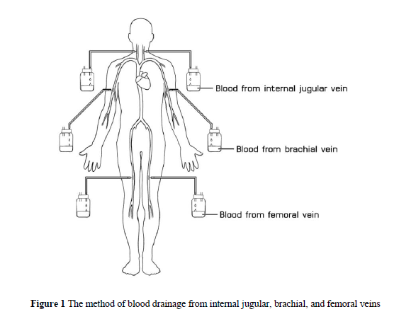
Two-point injections of embalming fluid without venous drainage, along with immersion in a post-fixative pool, has been used for several decades in the Department of Anatomy, Chiang Mai University (CMU). However, tissue decomposition was frequently observed during dissection. To address this, a Modified Embalming Method (MEM), utilizing a six-point injection technique with venous drainage, was tested. This study evaluates the effectiveness of MEM compared to the traditional two-point method with immersion. Ten cadavers were preserved and assessed for range of motion (ROM), histological integrity, and dissection quality. The cadavers were divided into two groups: five were embalmed using MEM and stored in plastic bags at room temperature, while the other five were preserved using the Present Embalming Method (PEM), which involved two-point injection without venous drainage followed by immersion. Both groups received the same embalming fluid. After one year, ROM was measured, and dissection quality was evaluated by ten dissectors. The MEM group showed greater joint mobility and superior tissue quality for both gross anatomical and histological analysis. The enhanced perfusion achieved by MEM ensured uniform distribution of fixative throughout the body. Furthermore, MEM eliminated the need for immersion, reduced chemical use, and allowed safe storage of cadavers in mortuary bags at room temperature.
References
Ajileye, A. B., Esan, E. O., & Adeyemi, O. A. (2018). Human embalming techniques: a review. American Journal of Biomedical Sciences, 10(2), 82-95. https://doi.org/10.5099/aj180200082
Alsharif, M. H. K., Musthafa, M., Elamin, A. Y., Ibnouf, E. O., Taha, K. M., Alfaki, M. A., ... & Aldosari, K. H. M. (2017). A brief review on the principles of human cadaver preservation and monitoring of microbial degradation. Forensic Medicine and Anatomy Research, 5(3), 73–83. https://doi.org/10.4236/fmar.2017.53003
Anderson, S. D. (2006). Practical light embalming technique for use in the surgical fresh tissue dissection laboratory. Clinical Anatomy, 19(1), 8-11. https://doi.org/10.1002/ca.20216
Balta, J. Y., Cronin, M., Cryan, J. F., & O'mahony, S. M. (2015a). Human preservation techniques in anatomy: a 21st century medical education perspective. Clinical Anatomy, 28(6), 725-734. https://doi.org/10.1002/ca.22585
Balta, J. Y., Lamb, C., & Soames, R. W. (2015b). A pilot study comparing the use of Thiel‐and formalin‐embalmed cadavers in the teaching of human anatomy. Anatomical Sciences Education, 8(1), 86-91. https://doi.org/10.1002/ase.1470
Barton, D. P., Davies, D. C., Mahadevan, V., Dennis, L., Adib, T., Mudan, S., ... & Ellis, H. (2009). Dissection of soft-preserved cadavers in the training of gynaecological oncologists: report of the first UK workshop. Gynecologic Oncology, 113(3), 352-356. https://doi.org/10.1016/j.ygyno.2009.02.012
Batra, A. P. S., Khurana, B. S., Mahajan, A., & Kaur, N. (2010). Embalming and other methods of dead body preservation. International Journal of Medical Toxicology and Legal Medicine, 12(3), 15–19.
Bertone, V. H., Blasi, E., Ottone, N. E., & Dominguez, M. L. (2011). Walther Thiel method for the preservation of corpses with maintenance of the main physical properties of vivo. Revista Argentina de Anatomía, 2(3), 71–100.
Bradbury, S. A., & Hoshino, K. (1978). An improved embalming procedure for long-lasting preservation of the cadaver for anatomical study. Cells Tissues Organs, 101(2), 97-103. https://doi.org/10.1159/000144954
Brenner, E. (2014). Human body preservation–old and new techniques. Journal of anatomy, 224(3), 316-344. https://doi.org/10.1111/joa.12160
Burn, C. G. (1934). Experimental studies of postmortem bacterial invasion in animals. The Journal of Infectious Diseases, 54(3), 388-394. https://doi.org/10.1093/infdis/54.3.388
Chen, Y., Bundy, D. S., & Hoff, S. J. (1999). Using olfactometry to measure intensity and threshold dilution ratio for evaluating swine odor. Journal of the Air & Waste Management Association, 49(7), 847-853. https://doi.org/10.1080/10473289.1999.10463855
De Varrimento, E. (2013). Improvement of the embalming perfusion method: The innovation and the results by light and scanning electron microscopy. Acta Médica Portuguesa, 26(3), 188–194.
Durongphan, A., Chongkolwatana, W., Ngamskulrungroj, P., Pochnasomboon, T., Pinkaew, J., Pamornpol, B., ... & Ongsiriporn, M. (2022). A pilot comparative study of submerged vs. non-submerged saturated salt solution human cadavers embalming method by gross, histological, and microbiological evaluation. Siriraj Medical Journal, 74(7), 431–439. https://doi.org/10.33192/Smj.2022.52
Eisma, R., Lamb, C., & Soames, R. W. (2013). From formalin to Thiel embalming: What changes? One anatomy department's experiences. Clinical Anatomy, 26(5), 564–571. https://doi.org/10.1002/ca.22222
Fox, C. H., Johnson, F. B., Whiting, J., & Roller, P. P. (1985). Formaldehyde fixation. Journal of Histochemistry and Cytochemistry, 33(8), 845–853. https://doi.org/10.1177/33.8.3894502
Hammer, N., Löffler, S., Bechmann, I., Steinke, H., Hädrich, C., & Feja, C. (2015). Comparison of modified thiel embalming and ethanol‐glycerin fixation in an anatomy environment: Potentials and limitations of two complementary techniques. Anatomical Sciences Education, 8(1), 74-85. https://doi.org/10.1002/ase.1450
Hammer, N., Löffler, S., Feja, C., Sandrock, M., Schmidt, W., Bechmann, I., & Steinke, H. (2012). Ethanol‐glycerin fixation with thymol conservation: a potential alternative to formaldehyde and phenol embalming. Anatomical Sciences Education, 5(4), 225-233. https://doi.org/10.1002/ase.1270
Hayashi, S., Homma, H., Naito, M., Oda, J., Nishiyama, T., Kawamoto, A., ... & Itoh, M. (2014). Saturated salt solution method: a useful cadaver embalming for surgical skills training. Medicine, 93(27), Article e196. https://doi.org/10.1097/MD.0000000000000196
Healy, S. E., Rai, B. P., Biyani, C. S., Eisma, R., Soames, R. W., & Nabi, G. (2015). Thiel embalming method for cadaver preservation: a review of new training model for urologic skills training. Urology, 85(3), 499-504. https://doi.org/10.1016/j.urology.2014.11.009
Heimesaat, M. M., Boelke, S., Fischer, A., Haag, L. M., Loddenkemper, C., Kühl, A. A., ... & Bereswill, S. (2012). Comprehensive postmortem analyses of intestinal microbiota changes and bacterial translocation in human flora associated mice. PLoS One, 7(7), Article e40758. https://doi.org/10.1371/journal.pone.0040758
Kaliappan, A., Motwani, R., Gupta, T., & Chandrupatla, M. (2023). Innovative cadaver preservation techniques: a systematic review. Maedica, 18(1), 127-135. https://doi.org/10.26574/maedica.2023.18.1.127
Kellerma, G. D., Waterman, N. G., & Scharfenberger, L. F. (1976). Demonstration in vitro of postmortem bacterial transmigration. American Journal of Clinical Pathology, 66(5), 911-915. https://doi.org/10.1093/ajcp/66.5.911
Kerckaert, I., Van Hoof, T., Pattyn, P., & D’Herde, K. (2008). Endogent: Centre for anatomy and invasive techniques. Anatomy, 2(1), 28–33.
Mayer, R. G. (2011). Embalming: History, theory, and practice (5th ed.). New York, NY: McGraw-Hill.
Mills, P. R. (2010). Preparation and presentation of anatomical specimens at the University of Sydney. Anatomy Department, University of Sydney. http://fliphtml5.com/mlay/rbhb/basic
Natekar, P. E., & Desouza, F. M. (2012). A new embalming fluid for preserving cadavers. Journal of Krishna Institute of Medical Sciences University, 1(2), 76-80.
Riedel, S. (2014). The value of postmortem microbiology cultures. Journal of Clinical Microbiology, 52(4), 1028-1033. https://doi.org/10.1128/jcm.03102-13
Sugata, Y., Miyaso, H., Odaka, Y., Komiyama, M., Sakamoto, N., Mori, C., & Matsuno, Y. (2016). Levels of formaldehyde vapor released from embalmed cadavers in each dissection stage. Environmental Science and Pollution Research, 23, 16176-16182. https://doi.org/10.1007/s11356-016-6744-8
Thavarajah, R., Mudimbaimannar, V. K., Elizabeth, J., Rao, U. K., & Ranganathan, K. (2012). Chemical and physical basics of routine formaldehyde fixation. Journal of Oral and Maxillofacial Pathology, 16(3), 400-405. https://doi.org/10.4103/0973-029X.102496
Thaweekhotr, P., Thongsopha, C., Tanprawate, S., Sudwan, P., Bumroongkit, K., & Quiggins, R. (2022). Incidence of death from stroke and brains of stroke victim cadavers in Northern Thailand. Chiang Mai University Journal of Natural Sciences, 21(2), Article e2022028.
Thaweekhotr, P., Thongsopha, C., Sudwan, P., Bumroongkit, K., Seeharach, K., Jaiyen, C., Suannahoy, P., Phasukdee, N., & Quiggins, R. (2023). Histological features of cerebral arteries and brain parenchyma in cadavers who had died from hemorrhagic stroke. Journal of Current Science and Technology, 13(3), 574–583. https://doi.org/10.59796/jcst.V13N3.2023.768
Theeuwes, H. P., van Riel, M. P. J. M., Lange, J. F., & Kleinrensink, G. J. (2017). A new model for training on human specimens in surgical-anatomical skills labs. Anatomy Physiology and Biochemistry International Journal, 3(1), 1–5. https://doi.org/10.19080/APBIJ.2017.03.555604
Thiel, W. (1992). Die Konservierung ganzer Leichen in natürlichen Farben. Annals of Anatomy – Anatomischer Anzeiger, 174(3), 185–195. https://doi.org/10.1016/S0940-9602(11)80346-8
Thiel, W. (2002). Ergänzung für die Konservierung ganzer Leichen nach W. Thiel. Annals of Anatomy – Anatomischer Anzeiger, 184(3), 267–269. https://doi.org/10.1016/S0940-9602(02)80121-2
Tolhurst, D. E., & Hart, J. (1990). Cadaver preservation and dissection. European Journal of Plastic Surgery, 13, 75-78. https://doi.org/10.1007/BF00177811
Downloads
Published
How to Cite
Issue
Section
License
Copyright (c) 2025 Journal of Current Science and Technology

This work is licensed under a Creative Commons Attribution-NonCommercial-NoDerivatives 4.0 International License.