Clinical and Optical Coherence Tomography Characteristics of Large Primary Macular Holes
DOI:
https://doi.org/10.59796/jcst.V16N4.2026.223Keywords:
ellipsoid zone, external limiting membrane, large primary macular hole, macular hole indices, optical coherence tomography, visual acuity, vitreomacular interfaceAbstract
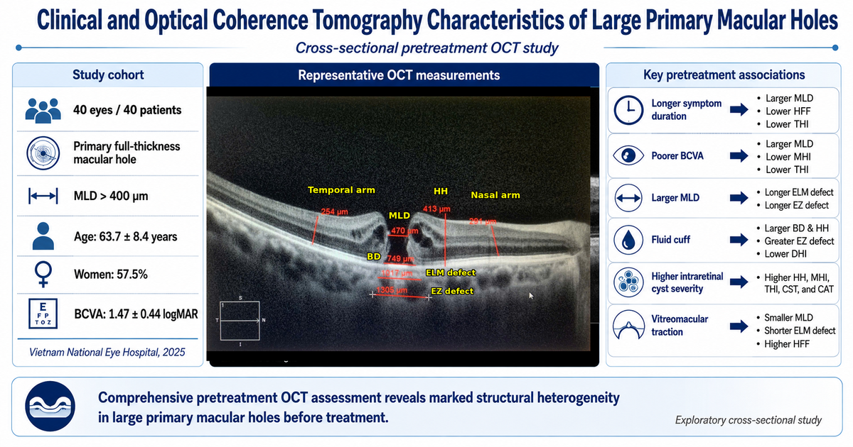
Large primary full-thickness macular holes are associated with visual impairment and complex optical coherence tomography (OCT) abnormalities. This single-center cross-sectional study described pretreatment clinical and OCT characteristics of large primary macular holes and explored associations with symptom duration and visual acuity. Forty eyes from 40 surgical candidates with primary macular holes and a minimum linear diameter (MLD) > 400 µm were analyzed at Vietnam National Eye Hospital, Hanoi, Vietnam, from March to December 2025. Clinical variables included age, sex, symptom duration, and best-corrected visual acuity (BCVA). OCT variables included vitreomacular status, fluid cuff, intraretinal cyst severity, MLD, base diameter, hole height, external limiting membrane (ELM) and ellipsoid zone (EZ) defect lengths, macular thickness parameters, and macular hole indices. Mean age was 63.7 ± 8.4 years, and women accounted for 57.5% of patients. Mean BCVA was 1.47 ± 0.44 logMAR. Mean MLD, base diameter, and hole height were 594.3 ± 155.8 µm, 1234.9 ± 456.3 µm, and 480.3 ± 110.4 µm, respectively. Complete posterior vitreous detachment, fluid cuff, and intraretinal cysts of any severity were observed in 77.5%, 57.5%, and 85.0% of eyes, respectively. Longer symptom duration was associated with larger MLD and lower HFF and THI. Poorer BCVA was associated with larger MLD and lower MHI and THI. Outer retinal defect lengths correlated with hole size, and fluid cuff was associated with wider structural disruption. Comprehensive pretreatment OCT assessment helps characterize structural severity in large primary macular holes. These associations are exploratory and should be validated prospectively.
References
Ch'ng, S. W., Patton, N., Ahmed, M., Ivanova, T., Baumann, C., Charles, S., & Jalil, A. (2018). The Manchester large macular hole study: Is it time to reclassify large macular holes?. American Journal of Ophthalmology, 195, 36-42. https://doi.org/10.1016/j.ajo.2018.07.027
Duker, J. S., Kaiser, P. K., Binder, S., de Smet, M. D., Gaudric, A., Reichel, E., ... & Stalmans, P. (2013). The International Vitreomacular Traction Study Group classification of vitreomacular adhesion, traction, and macular hole. Ophthalmology, 120(12), 2611-2619. https://doi.org/10.1016/j.ophtha.2013.07.042
Forsaa, V. A., Lindtjørn, B., Kvaløy, J. T., Frøystein, T., & Krohn, J. (2018). Epidemiology and morphology of full‐thickness macular holes. Acta Ophthalmologica, 96(4), 397-404. https://doi.org/10.1111/aos.13618
Gass, J. D. M. (1988). Idiopathic senile macular hole: Its early stages and pathogenesis. Archives of Ophthalmology, 106(5), 629-639. https://doi.org/10.1001/archopht.1988.01060130683026
Goto, K., Iwase, T., Yamamoto, K., Ra, E., & Terasaki, H. (2020). Correlations between intraretinal cystoid cavities and pre-and postoperative characteristics of eyes after closure of idiopathic macular hole. Scientific Reports, 10(1), Article 2310. https://doi.org/10.1038/s41598-020-59295-7
Houly, J. R., Veloso, C. E., Passos, E., & Nehemy, M. B. (2017). Quantitative analysis of external limiting membrane, ellipsoid zone and interdigitation zone defects in patients with macular holes. Graefe's Archive for Clinical and Experimental Ophthalmology, 255(7), 1297-1306. https://doi.org/10.1007/s00417-017-3636-7
Jenchitr, W., Yokkampon, P., Ploysit, P., & Ausayakhun, S. (2024). The prevalence of visual impairment of the Elderly at University Eye Clinic. Journal of Current Science and Technology, 14(1), Article 9. https://doi.org/10.59796/jcst.V14N1.2024.9
Kusuhara, S., Escaño, M. F. T., Fujii, S., Nakanishi, Y., Tamura, Y., Nagai, A., ... & Negi, A. (2004). Prediction of postoperative visual outcome based on hole configuration by optical coherence tomography in eyes with idiopathic macular holes. American Journal of Ophthalmology, 138(5), 709-716. https://doi.org/10.1016/j.ajo.2004.04.063
La Cour, M., & Friis, J. (2002). Macular holes: Classification, epidemiology, natural history and treatment. Acta Ophthalmologica Scandinavica, 80(6), 579-587. https://doi.org/10.1034/j.1600-0420.2002.800605.x
Murphy, D. C., Al-Zubaidy, M., Lois, N., Scott, N., Steel, D. H., Qu, J., ... & Alvin, K. K. H. (2023). The effect of macular hole duration on surgical outcomes: An individual participant data study of randomized controlled trials. Ophthalmology, 130(2), 152-163. https://doi.org/10.1016/j.ophtha.2022.08.028
Oh, J., Smiddy, W. E., Flynn, H. W., Gregori, G., & Lujan, B. (2010). Photoreceptor inner/outer segment defect imaging by spectral domain OCT and visual prognosis after macular hole surgery. Investigative Ophthalmology & Visual Science, 51(3), 1651-1658. https://doi.org/10.1167/iovs.09-4420
Ozturk, Y., Ağın, A., Kockar, N., Ay, N., Imamoglu, S., Ozcelik Kose, A., & Kugu, S. (2022). The importance of anatomic configuration and cystic changes in macular hole: Predicting surgical success with a different approach. Current Eye Research, 47(10), 1436-1443. https://doi.org/10.1080/02713683.2022.2096908
Radke, N. V., Ruamviboonsuk, P., Steel, D. H., Tian, T., Hunyor, A. P., Tsai, A. S., ... & Lam, D. S. (2025). Controversies, consensuses, and guidelines on macular hole surgery by the Asia–Pacific Vitreo-retina Society (APVRS) and the Asia–Pacific Academy of Professors in Ophthalmology (AAPPO). Eye and Vision, 12(1), Article 30. https://doi.org/10.1186/s40662-025-00446-0
Rezende, F. A., Ferreira, B. G., Rampakakis, E., Steel, D. H., Koss, M. J., Nawrocka, Z. A., ... & Duker, J. S. (2023). Surgical classification for large macular hole: Based on different surgical techniques results: The CLOSE study group. International Journal of Retina and Vitreous, 9(1), Article 4. https://doi.org/10.1186/s40942-022-00439-4
Ruiz-Moreno, J. M., Staicu, C., Piñero, D. P., Montero, J., Lugo, F., & Amat, P. (2008). Optical coherence tomography predictive factors for macular hole surgery outcome. British Journal of Ophthalmology, 92(5), 640-644. https://doi.org/10.1136/bjo.2007.136176
Sinawat, S., Jumpawong, S., Ratanapakorn, T., Bhoomibunchoo, C., Yospaiboon, Y., & Sinawat, S. (2021). Efficacy of pars plana vitrectomy with internal limiting membrane peeling for treatment of large idiopathic full-thickness macular holes. Clinical Ophthalmology, 15, 521-529. https://doi.org/10.2147/OPTH.S294190
The Royal College of Ophthalmologists. (2025). Idiopathic full thickness macular holes: Clinical guideline. Retrieved from https://www.rcophth.ac.uk/resources-listing/idiopathic-full-thickness-macular-holes/
Venkatesh, R., Mohan, A., Sinha, S., Aseem, A., & Yadav, N. K. (2019). Newer indices for predicting macular hole closure in idiopathic macular holes: A retrospective, comparative study. Indian Journal of Ophthalmology, 67(11), Article 1857. https://doi.org/10.4103/ijo.IJO_364_19
Yang, J., Xia, H., Liu, Y., Wang, X., Yuan, H., Hou, Q., ... & Li, X. (2021). Ellipsoid zone and external limiting membrane-related parameters on spectral domain-optical coherence tomography and their relationships with visual prognosis after successful macular hole surgery. Frontiers in Medicine, 8, Article 779602. https://doi.org/10.3389/fmed.2021.779602
Yun, C., Oh, J., Hwang, S. Y., Togloom, A., Kim, S. W., & Huh, K. (2012). Morphologic characteristics of chronic macular hole on optical coherence tomography. Retina, 32(10), 2077-2084. https://doi.org/10.1097/IAE.0b013e31825620ba
Downloads
Published
How to Cite
License
Copyright (c) 2026 Journal of Current Science and Technology

This work is licensed under a Creative Commons Attribution-NonCommercial-NoDerivatives 4.0 International License.