Diagnostic Accuracy of Cybersight AI for Glaucoma Detection: Sensitivity, Specificity, and ROC Analysis
DOI:
https://doi.org/10.59796/jcst.V16N4.2026.220Keywords:
artificial intelligence, deep learning, diagnostic accuracy, glaucoma, sensitivity and specificityAbstract
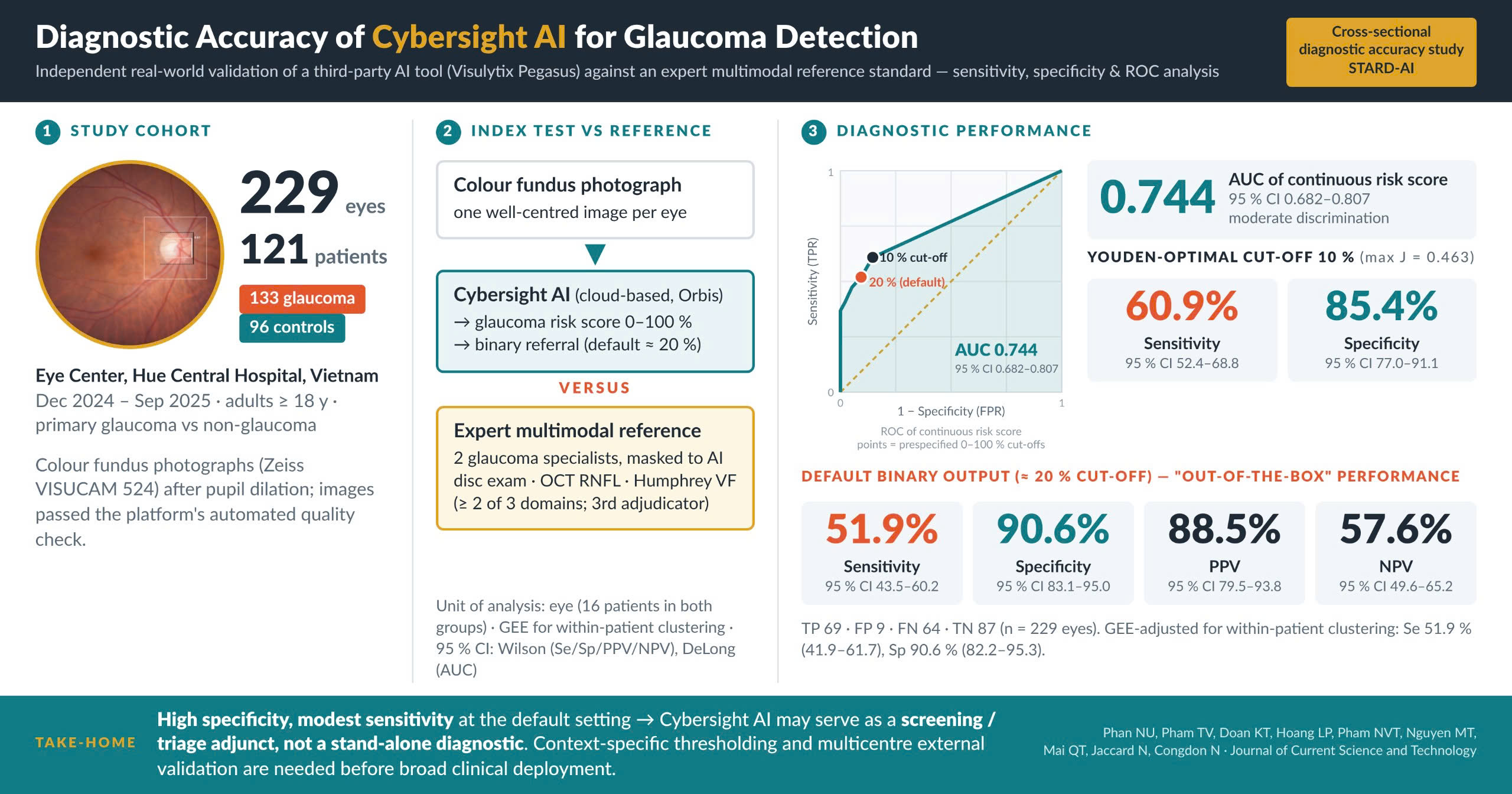
Glaucoma is a chronic, progressive optic neuropathy and a leading cause of irreversible blindness worldwide. Early detection is crucial but is often limited by nonspecific clinical signs and the need for specialist expertise. Artificial intelligence (AI), particularly deep learning, has shown promise for glaucoma detection from ophthalmic images. This study independently validated an existing third-party AI tool, Cybersight AI, powered by the Visulytix Pegasus engine, for detecting primary glaucoma. No new algorithm was developed or proposed. A cross-sectional diagnostic accuracy study was conducted on 229 eyes from 121 patients, including 133 eyes with primary glaucoma and 96 controls, at Hue Central Hospital between December 2024 and September 2025. Fundus photographs were analyzed using the Cybersight AI platform, which estimated the vertical cup-to-disc ratio and generated a binary glaucoma referral recommendation. AI outputs were compared with diagnoses established by ophthalmologists using an expert multimodal clinical reference standard. Diagnostic performance was assessed using sensitivity, specificity, positive and negative predictive values, and the area under the receiver operating characteristic curve (AUC). At its default operating point, Cybersight AI achieved 51.9% sensitivity (95% CI 43.5–60.2) and 90.6% specificity (95% CI 83.1–95.0), with an AUC of 0.744 (95% CI 0.682–0.807). Positive and negative predictive values were 88.5% and 57.6%, respectively. The Youden-optimal cut-off was 10%, yielding 60.9% sensitivity and 85.4% specificity. Cybersight AI demonstrated moderate discrimination, high specificity, and modest sensitivity at its default binary setting in this Vietnamese tertiary-care cohort. It may serve as a screening or triage adjunct, although context-specific thresholding and further external validation are required before broad clinical deployment. Limitations include the single-center convenience sample, within-patient clustering, and comparison of a fundus-only index test against a multimodal reference standard. Future studies should prioritize multicenter validation in Vietnamese settings and comparison with expert fundus assessment alone.
References
Anton, N., Doroftei, B., Curteanu, S., Catãlin, L., Ilie, O. D., Târcoveanu, F., & Bogdănici, C. M. (2023). Comprehensive review on the use of artificial intelligence in ophthalmology and future research directions. Diagnostics, 13(1), Article 100. https://doi.org/10.3390/diagnostics13010100
Ashtari-Majlan, M., Dehshibi, M. M., & Masip, D. (2024). Glaucoma diagnosis in the era of deep learning: A survey. Expert Systems with Applications, 256, Article 124888. https://doi.org/10.1016/j.eswa.2024.124888
Budenz, D. L., Rhee, P., Feuer, W. J., McSoley, J., Johnson, C. A., & Anderson, D. R. (2002). Comparison of glaucomatous visual field defects using standard full threshold and Swedish interactive threshold algorithms. Archives of Ophthalmology, 120(9), 1136-1141. https://doi.org/10.1001/archopht.120.9.1136
Chaurasia, A. K., Greatbatch, C. J., & Hewitt, A. W. (2022). Diagnostic accuracy of artificial intelligence in glaucoma screening and clinical practice. Journal of Glaucoma, 31(5), 285-299. https://doi.org/10.1097/IJG.0000000000002015
Chuter, B., Huynh, J., Bowd, C., Walker, E., Rezapour, J., Brye, N., ... & Christopher, M. (2024). Deep learning identifies high-quality fundus photographs and increases accuracy in automated primary open angle glaucoma detection. Translational Vision Science & Technology, 13(1), Article 23. https://doi.org/10.1167/tvst.13.1.23
Crowston, J. G., Hopley, C. R., Healey, P. R., Lee, A., & Mitchell, P. (2004). The effect of optic disc diameter on vertical cup to disc ratio percentiles in a population based cohort: The Blue Mountains Eye Study. British Journal of Ophthalmology, 88(6), 766-770. https://doi.org/10.1136/bjo.2003.028548
Foster, P. J., Buhrmann, R., Quigley, H. A., & Johnson, G. J. (2002). The definition and classification of glaucoma in prevalence surveys. British Journal of Ophthalmology, 86(2), 238-242. https://doi.org/10.1136/bjo.86.2.238
Guni, A., Sounderajah, V., Whiting, P., Bossuyt, P., Darzi, A., & Ashrafian, H. (2024). Revised tool for the quality assessment of diagnostic accuracy studies using AI (QUADAS-AI): protocol for a qualitative study. JMIR Research Protocols, 13(1), Article e58202. https://doi.org/10.2196/58202
Jaccard, N. (2022). AI for glaucoma care. Retrieved from https://glaucomatoday.com/articles/2022-july-aug/ai-for-glaucoma-care
Jenchitr, W., Yokkampon, P., Ploysit, P., & Ausayakhun, S. (2024). The prevalence of visual impairment of the Elderly at University Eye Clinic. Journal of Current Science and Technology, 14(1), Article 9. https://doi.org/10.59796/jcst.V14N1.2024.9
Jin, Y., Liang, L., Li, J., Xu, K., Zhou, W., & Li, Y. (2024). Artificial intelligence and glaucoma: A lucid and comprehensive review. Frontiers in Medicine, 11, Article 1423813. https://doi.org/10.3389/fmed.2024.1423813
La Bruna, S., Rai, A., Mao, G., Kerr, J., Amin, H., Zemborain, Z. Z., ... & Hood, D. C. (2022). The OCT RNFL probability map and artifacts resembling glaucomatous damage. Translational Vision Science & Technology, 11(3), Article 18. https://doi.org/10.1167/tvst.11.3.18
Lan, C. H., Chiu, T. H., Yen, W. T., & Lu, D. W. (2025). Artificial intelligence in glaucoma: Advances in diagnosis, progression forecasting, and surgical outcome prediction. International Journal of Molecular Sciences, 26(10), Article 4473. https://doi.org/10.3390/ijms26104473
Ling, X. C., Chen, H. S. L., Yeh, P. H., Cheng, Y. C., Huang, C. Y., Shen, S. C., & Lee, Y. S. (2025). Deep learning in glaucoma detection and progression prediction: a systematic review and meta-analysis. Biomedicines, 13(2), Article 420. https://doi.org/10.3390/biomedicines13020420
Myers, J. G., Chen, T. C., Huang, D., & Schuman, J. S. (2024). Is it glaucoma or not? Watch for RNFL OCT artifacts. Retrieved from https://www.aao.org/eyenet/article/is-it-glaucoma-or-not-watch-for-rnfl-oct-artifacts
Qi, T., Liu, H., Frühn, L., Löw, K., Cursiefen, C., & Prokosch, V. (2025). Understanding glaucoma: Why it remains a leading cause of blindness worldwide. Klinische Monatsblätter Für Augenheilkunde, 242(07), 712-717. https://doi.org/10.1055/a-2617-1575
Ruia, S., & Tripathy, K. (2025). Humphrey visual field. StatPearls Publishing. Retrieved from https://pubmed.ncbi.nlm.nih.gov/36256759/
Senjam, S. S. (2020). Glaucoma blindness–A rapidly emerging non-communicable ocular disease in India: Addressing the issue with advocacy. Journal of Family Medicine and Primary Care, 9(5), Article 2200. https://doi.org/10.4103/jfmpc.jfmpc_111_20
Senthil, S., Rao, D. P., Savoy, F. M., Negiloni, K., Bhandary, S., Chary, R., & Chandrashekar, G. (2025). Evaluating real-world performance of an automated offline glaucoma AI on a smartphone fundus camera across glaucoma severity stages. PLoS One, 20(6), Article e0324883. https://doi.org/10.1371/journal.pone.0324883
Shahriari, M. H., Asadi, F., Moghaddasi, H., Roshanpour, A., Sharifipour, F., & Khorrami, Z. (2025). Applications of machine learning in glaucoma diagnosis based on tabular data: A systematic review. BMC Biomedical Engineering, 7(1), Article 9. https://doi.org/10.1186/s42490-025-00095-3
Shyamalee, T., Meedeniya, D., Lim, G., & Karunarathne, M. (2024). Automated tool support for glaucoma identification with explainability using fundus images. IEEE Access, 12, 17290-17307. https://doi.org/10.1109/ACCESS.2024.3359698
Soh, Z. D., Chee, M. L., Thakur, S., Tham, Y. C., Tao, Y., Lim, Z. W., ... & Cheng, C. Y. (2020). Asian‐specific vertical cup‐to‐disc ratio cut‐off for glaucoma screening: An evidence‐based recommendation from a multi‐ethnic Asian population. Clinical & Experimental Ophthalmology, 48(9), 1210-1218. https://doi.org/10.1111/ceo.13836
Sounderajah, V., Guni, A., Liu, X., Collins, G. S., Karthikesalingam, A., Markar, S. R., ... & Ashrafian, H. (2025). The STARD-AI reporting guideline for diagnostic accuracy studies using artificial intelligence. Nature Medicine, 31(10), 3283-3289. https://doi.org/10.1038/s41591-025-03953-8
Tello, A., Vazquez, E., Villamizar, S. J., Mejía-Salgado, G., Duarte-Bueno, L. M., Acuña, M. F., & Galvis, V. (2026). Diagnostic accuracy of the non-compliance of the ISNT rule in glaucoma: Systematic review and meta-analysis. European Journal of Ophthalmology, 36(2), 434–447. https://doi.org/10.1177/11206721251392629
Tonti, E., Tonti, S., Mancini, F., Bonini, C., Spadea, L., D’Esposito, F., ... & Zeppieri, M. (2024). Artificial intelligence and advanced technology in glaucoma: A review. Journal of Personalized Medicine, 14(10), Article 1062. https://doi.org/10.3390/jpm14101062
Wanichwecharungruang, B., Jaksataphorn, P., Yuttitham, K., Vanichvaranont, S., & Harncharoen, K. (2011). Optic disc area and diameter of the central retinal vein occlusion fellow eyes, determined by optical coherence tomography. Journal of the Medical Association of Thailand, 94(3), S76-S80.
Wolvaardt, E., & Hu, V. H. (2021). Update on glaucoma. Community Eye Health, 34(112), Article 31.
Wu, H., Wang, Y., Li, F., Liu, Z., & Shi, F. (2023). The national, regional, and global impact of glaucoma as reported in the 2019 Global Burden of Disease Study. Archives of Medical Science: AMS, 19(6), Article 1913. https://doi.org/10.5114/aoms/172929
Youden, W. J. (1950). Index for rating diagnostic tests. Cancer, 3(1), 32-35. https://doi.org/10.1002/1097-0142(1950)3:1%3C32::AID-CNCR2820030106%3E3.0.CO;2-3
Downloads
Published
How to Cite
License
Copyright (c) 2026 Journal of Current Science and Technology

This work is licensed under a Creative Commons Attribution-NonCommercial-NoDerivatives 4.0 International License.