
SiCal-CytoNet: A Stain-Invariant, Calibrated Multi-Scale Network for Trustworthy Cytology on SIPaKMeD
Keywords:
Pap-smear cytology, Optical-density, Brier loss, Calibration, Prototype-guided metric headAbstract
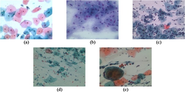
Background and Objectives: Cervical cancer screening through Pap-smear cytology remains a cornerstone of early diagnosis. However, its large-scale deployment is constrained by stain and illumination variability, morphological complexity across spatial scales, class imbalance, and unreliable confidence estimation in automated systems. Although deep learning models have achieved high classification accuracy on cytology benchmarks, many lack robustness to domain shifts and produce poorly calibrated probabilities, limiting their suitability for clinical triage. The objective of this research was to develop a stain-invariant, multi-scale, and well-calibrated deep learning framework that delivers both strong discrimination and trustworthy probability estimates for cervical cell classification, thereby enabling safe and effective decision support in screening workflows.
Methodology: This study proposes SiCal-CytoNet, a stain-aware and calibrated multi-scale network designed for Pap-smear cytology analysis. This framework employs optical-density–based stain deconvolution with controlled perturbations to address stain variability in combination with domain generalization strategies, including Fourier Domain Adaptation and MixStyle. Morphological features are learned at multiple resolutions and adaptively fused using a learnable resolution gate. A prototype-guided metric head improves class compactness under imbalance, while probability reliability is enhanced through a combination of Brier loss, evidential Dirichlet modeling, and post-hoc temperature scaling. A selective prediction mechanism based on calibrated confidence is integrated to support clinically meaningful abstention.
Main Results: On the SIPaKMeD five-class benchmark, SiCal-CytoNet achieves a test accuracy of 98.9% and a macro-F1 score of 98.7%, with excellent ranking performance reflected by an AUROC of 99.7% and an AUPRC of 99.4%. This model demonstrates strong probability calibration, attaining a low Brier score of 0.012 and an expected calibration error of 1.7%, while maintaining high discrimination under simulated stain, style, blur, and illumination shifts. Ablation experiments confirm the importance of multi-scale fusion, domain generalization, and calibration components. Using a utility-optimized selective prediction strategy, the system confidently automates 88.1% of cases while preserving high sensitivity, specificity, and predictive values for clinical triage.
Conclusions: The findings show that combining stain physics, adaptive multi-scale learning, prototype-guided classification, and explicit calibration produces a cytology model that is both highly accurate and clinically trustworthy. SiCal-CytoNet moves beyond accuracy-centric evaluation by delivering reliable probability estimates and robust performance under domain shifts, addressing critical requirements for real-world deployment in cervical cancer screening.
Practical Application: SiCal-CytoNet can be deployed as an automated triage system in cytology laboratories to assist pathologists by rapidly classifying routine Pap-smear samples while referring uncertain cases for expert review. Its calibrated outputs support transparent threshold selection, reduce screening workload, and enhance consistency across laboratories with heterogeneous staining conditions, offering tangible benefits to public health screening programs and AI-assisted diagnostic platforms.
References
Mathivanan, S.K., Francis, D., Srinivasan, S., Khatavkar, V.P.K. and Shah, M.A. 2024. Enhancing cervical cancer detection and robust classification through a fusion of deep learning models. Scientific Reports, 14, 10812.
Nour, M.K., Issaoui, I., Edris, A., Mahmud, A., Assiri, M. and Ibrahim, S.S. 2024. Computer aided cervical cancer diagnosis using gazelle optimization algorithm with deep learning model. IEEE Access, 12, 13046-13054.
Chikaraddi, A.K., Kanakaraddi, S., Kalwad, P., Kadole, A. and Budni, S. 2024. Cervical cancer diagnostics using machine learning algorithms. 5th International Conference for Emerging Technology (INCET), IEEE, May 2024, Belagavi, India, 1-6.
Singh, J., Kaliyar, R.K., Kumari, R., Sharma, N. and Prasad, P.D. 2024. Advancements in early detection of cervical cancer using machine learning and deep learning models for cervicography analysis. International Conference on Emerging Innovations and Advanced Computing (INNOCOMP), IEEE, May 2024, Bengaluru, India, 252-256.
Kumar, L.A., Jebarani, M.R.E. and Gokula Krishnan, V. 2023. Optimized deep belief neural network for semantic change detection in multi-temporal image. International Journal on Recent and Innovation Trends in Computing and Communication, 11, 86–93.
Sarhangi, H.A., Beigifard, D., Farmani, E. and Bolhasani, H. 2024. Deep learning techniques for cervical cancer diagnosis based on pathology and colposcopy images. Informatics in Medicine Unlocked, 47, 101503.
Pacal, I. 2024. MaxCerVixT: A novel lightweight vision transformer-based Approach for precise cervical cancer detection. Knowledge-Based Systems, 289, 111482.
Al Qathrady, M., Shaf, A., Ali, T., Farooq, U., Rehman, A., Alqhtani, S.M., Alshehri, M.S., Almakdi, S., Irfan, M., Rahman, S. and Eljak, L.A.B. 2024. A novel web framework for cervical cancer detection system: A machine learning breakthrough. IEEE Access, 12, 41542–41556.
Mehedi, M.H.K., Khandaker, M., Ara, S., Alam, M.A., Mridha, M.F. and Aung, Z. 2024. A lightweight deep learning method to identify different types of cervical cancer. Scientific Reports, 14, 29446.
Gokula Krishnan, V., Venkateswara Rao, P. and Divya, V. 2021. An energy efficient routing protocol based on SMO optimization in WSN. Proceedings of the 6th International Conference on Communication and Electronics Systems (ICCES), Coimbatore, India, 1040–1047.
Khare, S.K., Blanes‐Vidal, V., Booth, B.B., Petersen, L.K. and Nadimi, E.S. 2024. A systematic review and research recommendations on artificial intelligence for automated cervical cancer detection. Wiley Interdisciplinary Reviews: Data Mining and Knowledge Discovery, 14, e1550.
Wu, T., Lucas, E., Zhao, F., Basu, P. and Qiao, Y. 2024. Artificial intelligence strengthens cervical cancer screening–present and future. Cancer Biology & Medicine, 21, 864-879.
Baba, T., Miah, A.S.M., Shin, J. and Hasan, M.A.M. 2024. Cervical Cancer Detection using Multi-branch Deep Learning Model [Online], Available: https://arxiv.org/abs/2408.10498. [06 November 2025]
Xiong, L., Chen, C., Lin, Y., Song, Z. and Su, J. 2025. A Three-step automated segmentation method for early cervical cancer MRI images based on deep learning. International Journal of Imaging Systems and Technology, 35, e23207.
Krishnan, V.G., Rao, B.V.S., Prasad, J.R., Pushpa, P. and Kumari, S. 2024. Sugarcane yield prediction using NOA based swin transformer model in IoT smart agriculture. Journal of Applied Biology & Biotechnology, 12, 239–247.
Oak, P., Deshpande, P.S. and Iyer, B. 2025. Hybrid feature engineering for early prediction of cervical cancer using machine learning. Sensing and Imaging, 26, 39.
Paiboonborirak, C., Abu‐Rustum, N.R. and Wilailak, S. 2025. Artificial intelligence in the diagnosis and management of gynaecologic cancer. International Journal of Gynaecology & Obstetrics, 171, 199-209.
Provenzano, D., Wang, J., Goyal, S. and Rao, Y.J. 2025. Discussion of a simple method to generate descriptive images using predictive ResNet model weights and feature maps for recurrent cervix cancer. Tomography, 11, 38.
Navarajan, J., Jebarani, M.R.E. and Gokula Krishnan, V. 2023. Design of frequency reconfigurable antenna based on μC–μEMS switch. AEU-International Journal of Electronics and Communications, 171, 154911.
Chauhan, N.K., Singh, K., Kumar, A., Mishra, A., Gupta, S.K., Mahajan, S., Kadry, S. and Kim, J. 2025. A hybrid learning network with progressive resizing and PCA for diagnosis of cervical cancer on WSI slides. Scientific Reports, 15, 12801.
Downloads
Published
How to Cite
Issue
Section
License
Copyright (c) 2026 King Mongkut's University of Technology Thonburi

This work is licensed under a Creative Commons Attribution-NonCommercial-NoDerivatives 4.0 International License.
Any form of contents contained in an article published in Science and Engineering Connect, including text, equations, formula, tables, figures and other forms of illustrations are copyrights of King Mongkut's University of Technology Thonburi. Reproduction of these contents in any format for commercial purpose requires a prior written consent of the Editor of the Journal.

